Magnetic resonance imaging (MRI) is a medical imaging technique that produces detailed cross-sectional pictures of organs and soft tissues. This article explains what it is used for, which safety checks apply, the difference between scanner types and what happens during the exam.
What is an MRI scan and what is it used for?
MRI uses a strong magnetic field and radio waves to create images of the inside of the body; the computer can also build three-dimensional views (Mayo Clinic). It is mainly used to examine soft tissues such as the brain, spinal cord, joints, ligaments and abdominal or pelvic organs.
Doctors use it to diagnose conditions, look for the cause of symptoms and check whether a treatment is working, for example by following the size of a tumour (NHS). Typical requests include:
- Brain: to look for inflammation, vascular abnormalities or tumours.
- Spine: to assess discs, nerves and surrounding structures.
- Joints: to examine cartilage, tendons and ligaments.
- Abdomen and pelvis: to evaluate organs, blood vessels and lymph nodes (RadiologyInfo).
MRI is not automatically the first test. Whether it is the right exam, compared with X-ray, ultrasound or CT, depends on the clinical question and is decided by your doctor and the radiologist.
Does an MRI use radiation?
No. MRI does not use X-rays or other ionizing radiation; it relies on magnetic fields and radio energy (RadiologyInfo). When safety guidelines are followed, the exam carries almost no risk (RadiologyInfo).
The main hazard comes from the magnet itself: it can pull on metal objects and affect some implanted devices, which is why screening before the exam is strict. This is also why jewellery, watches and metal objects must stay outside the scanning room.
Who needs extra checks: implants, pregnancy and contrast
Implants and metal in the body
Pacemakers, implanted defibrillators, cochlear implants, some aneurysm clips, metal fragments and certain other devices can make an MRI unsafe or distort the images (Mayo Clinic). Some newer devices are acceptable, but the team must know the exact model, so bring any implant card or leaflet (RadiologyInfo). Orthopaedic metal generally poses no problem, though it can reduce image quality.
Pregnancy
No harmful effect has been reported since MRI came into use in the 1980s, but pregnant women are generally advised to avoid MRI in the first trimester unless the benefit clearly outweighs the risk (RadiologyInfo). Always tell the team if you are or might be pregnant or breastfeeding.
Contrast agent (gadolinium)
Some exams use a gadolinium-based contrast injected into a vein. It contains no iodine and allergic reactions are rare, but kidney function may be checked first, and people with kidney or liver disease or a previous contrast reaction should say so (RadiologyInfo). Contrast is usually avoided during pregnancy.
Closed or open MRI: what is the difference?
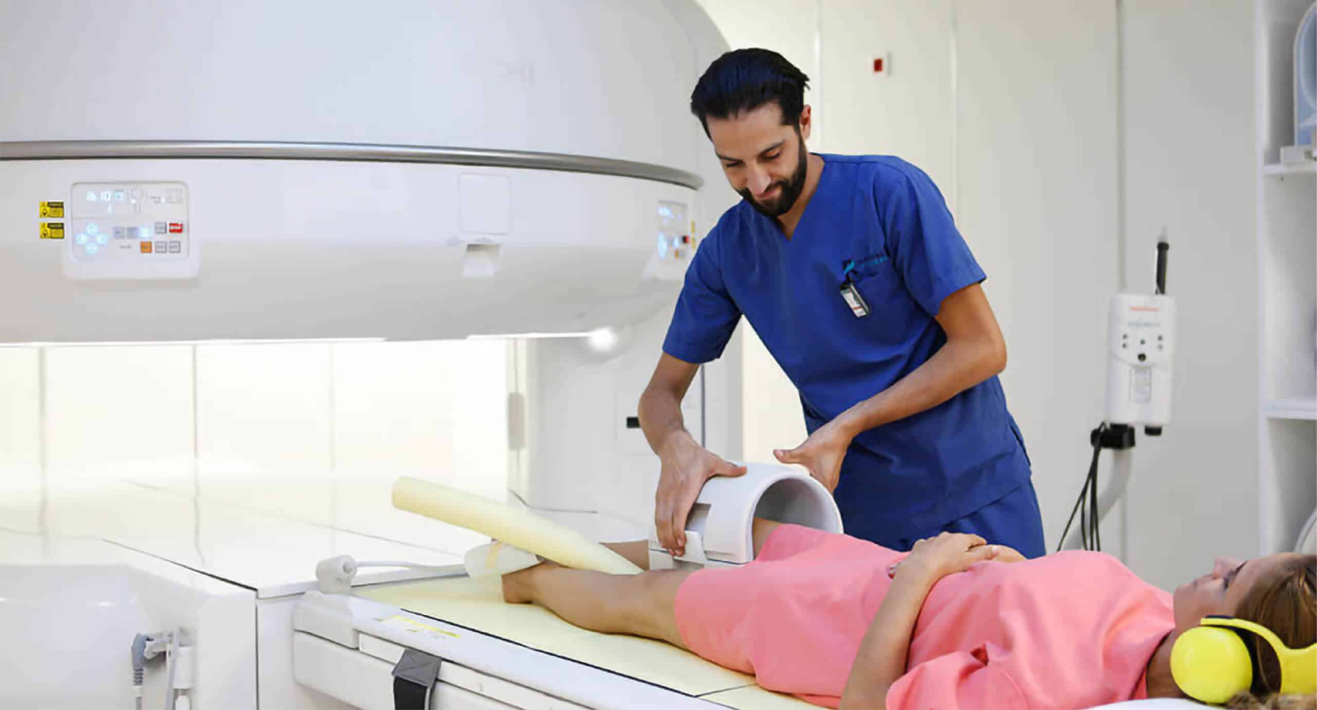
Most scanners are a tube open at both ends (Mayo Clinic). Their opening has a limited size and the table has a weight limit, so some large patients may not fit (RadiologyInfo). Some newer scanners have a wider opening for added comfort.
Open units, with more open sides, exist for some people who are very anxious or feel confined, but they are not available everywhere and are not suitable for every exam (RadiologyInfo). The choice of scanner depends on the examination requested and on what is available, so ask when booking.
How does the exam unfold?
Eating and drinking rules vary by exam and facility; follow the instructions you receive, and keep taking your usual medicines unless told otherwise (RadiologyInfo). Fasting is not needed for every MRI.
- You complete a safety questionnaire (implants, allergies, kidney problems, pregnancy).
- You change into a gown if needed and remove jewellery, watches, hairpins, hearing aids, removable dental work and other metal; leave bank cards and phones outside.
- You lie on a padded table that slides into the scanner.
- You are given earplugs or headphones, because the machine makes loud tapping and knocking sounds, and a squeeze ball to call the technologist, who can hear and speak to you throughout.
- You must stay still, as movement blurs the images.
The scan usually takes between 15 minutes and 1 hour, and sometimes longer (NHS); a typical complete visit is often quoted as 30 to 60 minutes. For children, a parent can often stay in the room after safety screening, and some children need preparation or sedation to stay still (RadiologyInfo).
Claustrophobia, anxiety and when to talk to your doctor
If you fear enclosed spaces, tell your doctor before the appointment. You may be offered a mild sedative, and you can ask to bring someone with you (NHS). If you take a sedative, do not drive yourself home (RadiologyInfo). Music, earplugs and the squeeze ball can also help, and the technologist can stop the scan at any time.
Speak to your doctor before booking if you have a pacemaker or any implant, if you are pregnant, if you have kidney disease or a contrast allergy, or if you are concerned about fitting in the scanner. The results are interpreted by a radiologist and explained by your referring doctor.
Further reading at La Rose Clinic
Frequently asked questions
Is an MRI painful or dangerous?
The exam is painless and uses no ionizing radiation. Risks are low when safety screening is followed, mainly concerning metal implants and, rarely, contrast reactions.
How long does an MRI take?
Usually between 15 minutes and 1 hour depending on the area and the exam, and sometimes longer.
Can I have an MRI with a pacemaker or metal implant?
Sometimes, depending on the exact device. Tell the team beforehand and bring your implant card so they can check whether it is MRI-compatible.
Do I need to fast before an MRI?
Not always. Rules vary by exam and facility, so follow the instructions you are given.
Can I have an MRI if I am claustrophobic?
Often yes. Tell your doctor in advance: a mild sedative, a wider scanner or an open unit may be options, depending on availability and the exam.
References
- MRI: Magnetic resonance imaging (Mayo Clinic) (mayoclinic.org)
- MRI scan (NHS) (nhs.uk)
- How to prepare for an MRI exam (RadiologyInfo.org) (radiologyinfo.org)
- Safety in MR imaging (RadiologyInfo.org) (radiologyinfo.org)
- Body MRI: abdomen, pelvis and chest (RadiologyInfo.org) (radiologyinfo.org)
This article is for information only. It does not replace a medical consultation: if in doubt, talk to your doctor.