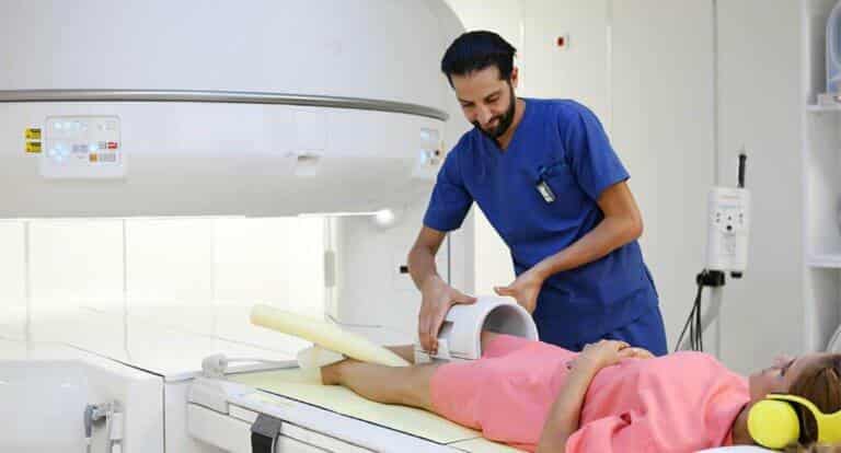

A mammogram does not always have the same purpose: it can look for breast cancer in a woman with no symptoms (screening) or investigate an abnormality that has already been noticed (diagnosis). This article explains that difference, the age guidelines, how to prepare, what happens during the exam and how to read a result.
Screening or diagnostic mammogram: what is the difference?
According to the National Cancer Institute, a screening mammogram looks for cancer before any symptoms appear, whereas a diagnostic mammogram is done after a lump or another change has been found by the patient or her doctor. Both use the same equipment, but the diagnostic exam takes images from more angles, which means a higher X-ray dose.
In France, the national programme promoted by the Institut national du cancer combines the mammogram with a clinical breast examination (looking at and feeling the breasts) carried out at the time of the exam (Santé.fr). If you have a symptom, you are therefore not in a screening situation: speak to a doctor first.
At what age should you be screened?
There is no universal age: recommendations vary by country, health system and level of risk.
- WHO: in settings with strong health systems, an organised mammogram every 2 years is recommended for women aged 50 to 69; before 50 and after 69, it is suggested only within a rigorous research and evaluation framework (WHO factsheet).
- France: an invitation every 2 years between the ages of 50 and 74 (Santé.fr).
- United States: the USPSTF task force recommends a mammogram every 2 years from age 40 to 74 for women at average risk (NCI).
- Tunisia: the Ministry of Health has presented a national screening programme; a surgeon quoted in the press advises a mammogram every 2 years from age 40, and every year if there is a family history of hereditary cancer (WebManagerCenter). This is a specialist’s opinion, not an official threshold.
People at higher risk, for example after chest radiotherapy in childhood or with a BRCA1 or BRCA2 mutation, may start earlier or be monitored more often (NCI). Your doctor will adapt the starting age and the frequency to your personal and family history.
How do you prepare for a mammogram?
Preparation is simple:
- On the day of the exam, avoid deodorant, antiperspirant, talc, powder, lotion, cream or perfume on the breasts and under the arms: these products can interfere with the reading of the images (NCI; NHS).
- Choose a top that is easy to remove (NHS).
- If possible, avoid scheduling the exam during or just before your period, when the breasts may be more tender (NCI).
If you are worried about pain, ask your doctor or pharmacist whether you could take a painkiller before the exam.
What happens during the exam?
Each breast is placed between two plates that compress it. In France, two images are taken of each breast and the compression lasts only a few seconds; it can be uncomfortable, or even painful, depending on the person (Santé.fr). According to the NHS, the mammogram itself takes only a few minutes and the whole appointment lasts no more than 30 minutes (NHS).
The radiologist may ask for extra images or an ultrasound, particularly if the breasts are dense. This does not necessarily mean that an abnormality has been found (Santé.fr).
Results: breast density and the BI-RADS classification
A first result is given after the exam. If no abnormality is seen, a second radiologist re-reads the images, and the final results are available in about 2 weeks. Out of 1,000 women screened, about 926 have no abnormality, 74 have one (most often benign or probably benign) and about 8 are diagnosed with cancer (Santé.fr).
The report usually includes a BI-RADS classification from 0 to 6 (NCI):
- 0: further imaging needed;
- 1: negative; 2: benign (continue routine screening);
- 3: probably benign (follow-up at 6 months);
- 4: suspicious (biopsy possible); 5: highly suggestive of cancer (biopsy needed);
- 6: cancer confirmed by biopsy.
Breast density describes the proportion of dense tissue and fatty tissue. In dense breasts, a mammogram is more likely to miss a cancer; this information is included in the report (NCI). Ask your radiologist what it means for your follow-up.
Warning signs and when to see a doctor
Do not wait for your next screening if you notice any of these changes (NHS):
- a lump or swelling in the breast, chest or armpit;
- dimpled skin (like orange peel or a dimple);
- nipple discharge, sometimes streaked with blood;
- a change in the shape or appearance of the nipple.
The NHS advises seeing a doctor about any change that is unusual for you. Knowing how your breasts normally look and feel helps you spot a change; this does not replace a clinical examination or the screening offered at your age. Your doctor, gynaecologist or midwife can examine your breasts and, depending on the situation, request a diagnostic mammogram.
Further reading at La Rose Clinic
Frequently asked questions
Does a mammogram hurt?
It can be uncomfortable, or even painful, depending on the person, because the breast is compressed between two plates; the compression lasts only a few seconds.
Can I wear deodorant on the day of the exam?
It is best to avoid it, as well as talc, powders and creams on the breasts and under the arms, because they can interfere with the reading of the images.
At what age should I have my first screening mammogram?
It depends on the recommendations in your country and your level of risk: 50 in France and in the WHO recommendations, 40 in the United States. Your doctor will adapt the starting age to your situation.
How long does the exam take?
The mammogram itself takes a few minutes; according to the NHS, the whole appointment lasts no more than 30 minutes.
What does a BI-RADS 0 result mean?
It means that further tests (extra images or an ultrasound) are needed before the mammogram can be classified; it is not a diagnosis of cancer.
References
- Dépistage du cancer du sein : l’essentiel du programme (INCa) (sante.fr)
- WHO position paper on mammography screening: factsheet (paho.org)
- Mammograms Fact Sheet (cancer.gov)
- What happens at your breast screening appointment (nhs.uk)
- Breast cancer: symptoms (nhs.uk)
- Octobre rose 2024 en Tunisie : programme national de dépistage (webmanagercenter.com)
This article is for information only. It does not replace a medical consultation: if in doubt, talk to your doctor.